
How Breast Arterial Calcification Signals a Higher Risk of Heart Disease in Women — and the Correct Follow-Up When It Appears on a Screening Mammogram
Chauncey W. Crandall IV, MD, FACC, FACP
Crandall Concierge Medicine & Cardiology
1411 N Flagler Dr., Suite 3902 · Farris Building · Good Samaritan Medical Center
West Palm Beach, Florida 33401
October 2026
Executive Summary
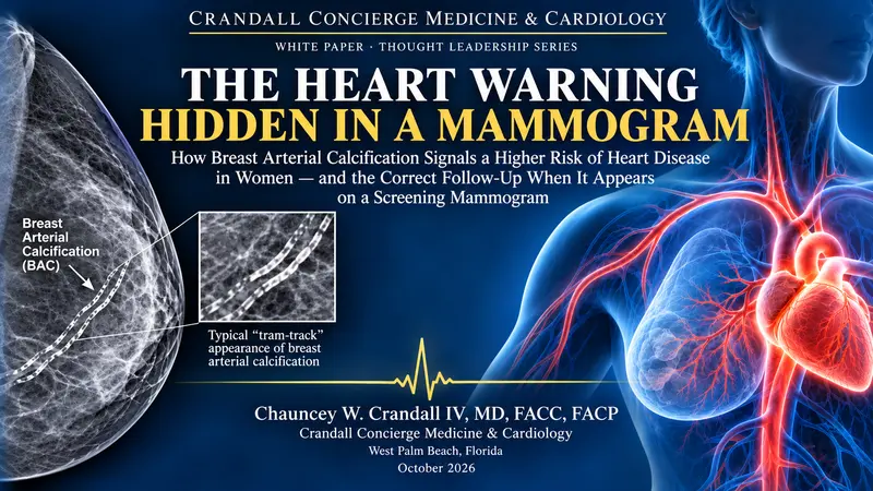
Heart disease remains the leading cause of death in women, yet the tools physicians use to estimate a woman’s risk routinely underestimate it.6,13 Millions of women already undergo a test every year that contains a second, largely ignored piece of cardiovascular information: the screening mammogram. Breast arterial calcification (BAC) — calcium deposits in the small arteries of the breast — is visible on a standard mammogram at no added cost, no added radiation, and no added appointment.1,6
A rapidly growing body of evidence, including several 2026 meta-analyses, shows that women with BAC have roughly twice the likelihood of coronary artery disease, about 1.8 times the rate of future cardiovascular events, and roughly double the risk of stroke, heart failure, and death from any cause.1,2 The added risk is most important in women whom conventional calculators label “low risk.”11,12
This paper reviews the evidence and makes a practical case: BAC should be treated as an additional, women-specific cardiovascular risk signal. It should not trigger stress tests or catheterization in a woman without symptoms, but it should trigger a structured cardiovascular evaluation. We present a stepwise follow-up pathway aligned with the 2026 ACC/AHA Multisociety Dyslipidemia Guideline, which now uses the PREVENT equations, universal lipoprotein(a) testing, and coronary artery calcium (CAC) scoring to refine treatment decisions.16
1. An Overlooked Signal in Women’s Heart Health
For decades, women’s cardiovascular disease has been underdiagnosed and undertreated. Women more often present with atypical symptoms, develop disease later, and are frequently classified as low risk by scores built largely on male-dominated cohorts. As a result, many women first learn they have heart disease when they suffer a heart attack or stroke.
Nearly two-thirds of American women age 40 and older have regular screening mammograms.4,15 Radiologists have long seen arterial calcium on these images and, historically, often left it out of the report because it has no bearing on breast cancer. That practice is changing. The American College of Radiology now supports reporting BAC, and leading reviews call for universal reporting paired with a conversation between patient and physician.6,15 Every mammogram report that mentions BAC is an opportunity for earlier prevention — if the finding is acted upon correctly.
2. What Breast Arterial Calcification Is
BAC appears on the mammogram as thin, parallel “tram-track” lines of calcium following the course of the breast arteries. It is distinct from the clustered microcalcifications that raise concern for cancer, and it has no relationship to breast cancer risk.
Pathologically, BAC is medial (Mönckeberg) calcification — calcium laid down in the muscular middle layer of the artery wall. This differs from the intimal, cholesterol-laden plaque that causes coronary blockages.6,10 The distinction matters: BAC is not simply “plaque in the breast.” It is a marker of arterial aging, stiffness, and metabolic stress that travels with coronary disease and predicts future events, even though it arises by a somewhat different mechanism.
BAC is common. It is found in roughly 10% to 17% of women undergoing screening, and its prevalence rises steeply with age.2,4 It is uncommon before menopause and increasingly frequent in women in their sixties and seventies, which is why the finding must always be interpreted in the context of the woman’s age.11
3. The Evidence Linking BAC to Heart Disease
Coronary artery disease already present
Pooled analyses consistently link BAC with existing coronary artery disease, with odds ratios of approximately 2.1 to 2.6, rising to about 3.5 in women without a prior diagnosis of coronary disease.3,4 A 2026 meta-analysis in the American Journal of Cardiology reported a pooled adjusted odds ratio of 4.00 (95% CI 2.44–6.56) for underlying coronary pathology.1
Future heart attacks, strokes, and death
The prospective data are equally compelling. A 2026 meta-analysis of approximately 25,000 women followed for 6 to 12 years found that BAC was associated with an 82% higher rate of incident cardiovascular events (pooled HR 1.82; 95% CI 1.37–2.43).1 A second, larger 2026 meta-analysis — 45 studies and 68,584 women — found BAC associated with roughly double the risk of stroke, heart failure, and all-cause mortality, and nearly triple the risk of cardiac death.2 In a 10-year prospective cohort, BAC-positive women had about three times the odds of developing coronary disease and five times the odds of stroke after adjustment for age.5
Table 1. Key evidence linking breast arterial calcification to cardiovascular disease
| Study | Population / Design | Key Finding |
| Adrejiya et al., Am J Cardiol 20261 | Meta-analysis; ~25,000 women; 6–12 yr follow-up | Incident CV events HR 1.82; coronary pathology OR 4.00 |
| Christensen et al., Am J Cardiol 20262 | Meta-analysis; 45 studies; 68,584 women | Stroke RR 2.05; heart failure RR 2.14; cardiac death RR 2.94; ASCVD RR 1.58; all-cause death RR 2.04 |
| Lee et al., PLoS One 20203; Osman et al., J Womens Health 20224 | Meta-analyses of prevalent coronary disease | CAD OR ~2.1–2.6; up to ~3.5 without prior CAD |
| Nudy et al., Menopause 20225 | 10-year prospective cohort | CAD OR 3.14; stroke OR 5.10 (age-adjusted) |
| Nerlekar et al., JACC Cardiovasc Imaging 202611 | Age-adjusted BAC percentile nomogram | Any BAC raised MACE in low-risk women; above-median BAC in intermediate/high-risk |
| Kim et al., ASCO 202614 | Retrospective; 650 women | Mean 10-yr ASCVD risk 14.0% with BAC vs 5.23% without |
4. Why BAC Is an Additional Risk Factor for Women
A finding earns its place as a clinical risk factor when it predicts outcomes beyond what we already measure. BAC meets that standard in three ways.
It tracks the metabolic drivers of disease. BAC is associated with diabetes (OR about 2.0), hypertension (OR about 1.8), and dyslipidemia.2,3 In the 2026 ASCO study, BAC-positive women carried nearly three times the average 10-year ASCVD risk of BAC-negative women.14
It adds information beyond traditional risk factors. Counterintuitively, BAC is inversely associated with smoking.2,3 That pattern confirms BAC is not merely a stand-in for the usual risk factors; it captures a different biology of arterial aging. Studies show BAC adds predictive value independent of traditional risk factors and is associated with progression of coronary atherosclerosis, and AI-quantified BAC has been shown in 2026 to predict cardiovascular morbidity and mortality.9,10
It matters most where current tools fail. The greatest incremental value of BAC is in women classified as low risk. In the 2026 age-percentile nomogram study, the presence of any BAC significantly increased major adverse cardiovascular events among low-risk women, while in intermediate- and high-risk women the risk rose with BAC above the age-adjusted median.11 Similar findings were reported in a 2026 primary-prevention cohort stratified by predicted risk.12 These are precisely the women who would otherwise be reassured and sent home.
Notably, the 2026 ACC/AHA guideline already recognizes several female-specific risk enhancers — early menopause, preeclampsia, gestational diabetes, and preterm delivery.16 BAC belongs in the same conceptual category: a finding unique to women that should move a clinician toward closer evaluation. Although no cardiovascular society guideline has yet formally incorporated BAC into its risk algorithm, the case for treating it as a risk-enhancing finding is strong.13
5. The Correct Follow-Up When BAC Is Present
The central principle is simple: a BAC finding should trigger a formal cardiovascular risk assessment, not an isolated reaction.7,8 In a woman without symptoms, BAC does not justify a stress test or a cardiac catheterization.8 Instead, it should launch the following stepwise evaluation.
Step 1 — Confirm and characterize the finding
Review the mammogram report for the presence and, where available, the severity of BAC (mild, moderate, or severe, or a quantitative or AI-derived score). Severity matters: the association with coronary disease is robust for moderate-to-severe BAC but less certain for mild BAC.3 Interpret the finding against the patient’s age, because BAC in a 50-year-old carries more weight than the same finding at 80.11
Step 2 — Perform a complete cardiovascular risk assessment
- Calculate 10-year and 30-year risk with the PREVENT-ASCVD equations, which replace the Pooled Cohort Equations in the 2026 ACC/AHA guideline.16
- Obtain a fasting or non-fasting lipid panel, lipoprotein(a) once in every adult, and ApoB when residual risk is suspected.16
- Measure blood pressure carefully, hemoglobin A1c, kidney function (eGFR and urine albumin-to-creatinine ratio), and body composition.
- Document women-specific and other risk enhancers: early menopause, preeclampsia, gestational diabetes, preterm delivery, autoimmune and inflammatory disease, and family history of premature heart disease.16
- Ask directly about exertional chest discomfort, unusual shortness of breath, fatigue, and palpitations. A woman with symptoms is evaluated for coronary disease on the basis of those symptoms — BAC simply adds urgency.
Step 3 — Act according to risk category
Table 2. Recommended follow-up of a BAC-positive woman without symptoms (aligned with the 2026 ACC/AHA Dyslipidemia Guideline)
| PREVENT 10-yr Risk | Recommended Next Step | Treatment Direction |
| High (≥10%) | No further imaging needed to start therapy. CAC optional for counseling. | Begin lipid-lowering therapy; LDL-C goal <70 mg/dL; control BP and glucose.16 |
| Intermediate (5% to <10%) | Obtain a CAC score — BAC strengthens the indication when the treatment decision is uncertain.7,16 | Therapy recommended; CAC result refines intensity and LDL-C goal. |
| Borderline (3% to <5%) | Treat BAC as a risk-enhancing finding; obtain a CAC score to guide shared decision-making. | Consider therapy, favored by BAC, other enhancers, or CAC >0. |
| Low (<3%) | Moderate–severe BAC, or BAC above the age-adjusted median, justifies a CAC score despite the low calculated risk.11 | Lifestyle optimization; therapy if CAC >0 or other enhancers present. Mild BAC: reassess. |
Step 4 — Interpret the CAC score
Table 3. CAC-guided management after a BAC finding (2026 ACC/AHA Dyslipidemia Guideline)16
| CAC Score (AU) | Management |
| 0 | Statin may generally be deferred unless diabetes, active smoking, familial hypercholesterolemia, or a strong family history is present. Continue lifestyle therapy and reassess periodically — BAC should not be forgotten. |
| 1–99 | Supports starting lipid-lowering therapy. |
| ≥100 | Lipid-lowering therapy indicated; LDL-C goal <70 mg/dL. |
| ≥300 | Consider a more aggressive LDL-C goal of <55 mg/dL. |
| ≥1,000 | Manage as a very-high-risk equivalent of established ASCVD; LDL-C goal <55 mg/dL. |
Step 5 — Pair every pathway with prevention and partnership
Regardless of category, a BAC finding should prompt lifestyle counseling — Mediterranean-style nutrition, regular aerobic and resistance exercise, sleep, weight management, and tobacco avoidance — along with tight control of blood pressure and blood sugar.7,8 Decisions about medication should be made through shared decision-making. Some clinicians may reasonably begin statin therapy without further testing given the strength of the BAC–cardiovascular association, though this is not guideline-mandated.7
What not to do
- Do not order stress testing or invasive angiography in a woman without symptoms solely because of BAC.8
- Do not assume a mammogram without BAC means low risk. BAC detects only about 23% to 63% of women who have coronary calcium, depending on the population, and every woman still needs standard risk assessment.8
- Do not ignore the finding. A BAC report that never reaches the patient’s physician is a missed opportunity for prevention.
6. Limitations and Unanswered Questions
Enthusiasm for BAC must be tempered by honest acknowledgment of its limits. Published meta-analyses show substantial heterogeneity among studies.2,3 There is no standardized method for grading or quantifying BAC, and no validated threshold for intervention, though AI-based scoring and age-percentile nomograms are rapidly closing that gap.10,11,13 Most importantly, no randomized outcome trial has yet shown that acting on BAC reduces heart attacks, strokes, or deaths.2,13 For now, BAC is a risk-refinement signal that prompts formal evaluation — not a stand-alone screening test or a diagnosis.
7. Implications for Clinical Practice
The practical opportunity is enormous and inexpensive. The image already exists; what is missing is a reliable pathway from the radiology report to the cardiology conversation. Practices can close that gap by asking breast imaging partners to report BAC routinely, preferably with a severity grade; by reviewing mammogram reports at every annual examination; and by building a standing protocol — PREVENT calculation, lipoprotein(a), and CAC scoring when indicated — for every BAC-positive patient.
In a concierge model, where the physician has the time to read the full mammogram report and the relationship to follow a woman across years, BAC becomes a powerful tool for personalized prevention. A finding that once went unmentioned can now be the moment a woman learns her heart needs attention — years before a heart attack makes the point for her.
Conclusion
Breast arterial calcification is a common, free, and underused marker of cardiovascular risk in women. The evidence now shows it is associated with roughly twice the risk of coronary disease, stroke, heart failure, and death, and that it adds the most information in the very women whom conventional scores reassure. BAC should be recognized as an additional risk factor for women. When it is present, the correct response is not alarm and not neglect, but a structured evaluation: a complete PREVENT-based risk assessment, lipoprotein(a) testing, CAC scoring when it will change management, guideline-directed lipid-lowering therapy, and a lifelong partnership in prevention.
References
- Adrejiya P, Bhanushali A, Mehta K, Amin M, Velarde GP. Breast arterial calcification on mammography and cardiovascular outcomes in women: a meta-analysis. Am J Cardiol. 2026;268:152-158. doi:10.1016/j.amjcard.2026.03.046
- Christensen E, Hillenbrand C, Kutty A, et al. A systematic review and meta-analysis of breast arterial calcification and its association with cardiovascular disease and all-cause mortality. Am J Cardiol. 2026;265:7-16. doi:10.1016/j.amjcard.2026.01.014
- Lee SC, Phillips M, Bellinge J, et al. Is breast arterial calcification associated with coronary artery disease? A systematic review and meta-analysis. PLoS One. 2020;15(7):e0236598. doi:10.1371/journal.pone.0236598
- Osman M, Regner S, Osman K, et al. Association between breast arterial calcification on mammography and coronary artery disease: a systematic review and meta-analysis. J Womens Health. 2022;31(12):1719-1726. doi:10.1089/jwh.2020.8733
- Nudy M, Asmaro R, Jiang X, Schnatz PF. The association between incidentally found breast arterial calcification on routine screening mammography and the development of coronary artery disease and stroke: results of a 10-year prospective study. Menopause. 2022;29(12):1375-1380. doi:10.1097/GME.0000000000002088
- Bui QM, Daniels LB. A review of the role of breast arterial calcification for cardiovascular risk stratification in women. Circulation. 2019;139(8):1094-1101. doi:10.1161/CIRCULATIONAHA.118.038092
- Quispe R, Al-Rifai M, Di Carlo PA, et al. Breast arterial calcium: a game changer in women’s cardiovascular health? JACC Cardiovasc Imaging. 2019;12(12):2538-2548. doi:10.1016/j.jcmg.2018.07.035
- Yoon YE, Yun B, Kim KM, Suh JW. Breast arterial calcification: a potential biomarker for atherosclerotic cardiovascular disease risk? Curr Atheroscler Rep. 2021;23(5):21. doi:10.1007/s11883-021-00924-5
- Yoon YE, Kim KM, Lee W, et al. Breast arterial calcification is associated with the progression of coronary atherosclerosis in asymptomatic women: a preliminary retrospective cohort study. Sci Rep. 2020;10(1):2755. doi:10.1038/s41598-020-59606-y
- Dapamede T, Urooj A, Joshi V, et al. Artificial intelligence–based quantification of breast arterial calcifications to predict cardiovascular morbidity and mortality. Eur Heart J. 2026;47(18):2206-2220. doi:10.1093/eurheartj/ehag128
- Nerlekar N, Soh CH, Vasanthakumar S, et al. A novel breast arterial calcification age-based percentile nomogram for the incremental prediction of incidental cardiovascular events. JACC Cardiovasc Imaging. 2026;19(6):735-748. doi:10.1016/j.jcmg.2026.03.008
- Ali TS, diFlorio-Alexander RM, Austin-Strohbehn JA, et al. Breast artery calcification for primary prevention of major adverse cardiovascular outcomes in women. JACC Adv. 2026;5(6 Pt 1):102737. doi:10.1016/j.jacadv.2026.102737
- Littman E, Bukhari S, Watts H, et al. Beyond the coronaries: utility of extra-coronary calcification for cardiovascular risk assessment in women. Curr Atheroscler Rep. 2026;28(1):57. doi:10.1007/s11883-026-01422-2
- Kim T, Shaw R, Abbate K, et al. The association between cardiovascular disease risk and breast arterial calcifications identified on screening mammography: a retrospective study. J Clin Oncol. 2026;44(16 Suppl):12026. doi:10.1200/JCO.2026.44.16_suppl.12026
- American College of Radiology. National Radiology Data Registry 2025. Reston, VA: American College of Radiology; 2025.
- Blumenthal RS, et al. 2026 ACC/AHA/AACVPR/ABC/ACPM/ADA/AGS/APhA/ASPC/NLA/PCNA Guideline on the Management of Dyslipidemia: a report of the American College of Cardiology/American Heart Association Joint Committee on Clinical Practice Guidelines. J Am Coll Cardiol. Published online March 13, 2026. doi:10.1016/j.jacc.2025.11.016
This white paper is provided for educational purposes and does not constitute individual medical advice. Patients should discuss their own mammogram findings and cardiovascular risk with their physician.